Condition
My legs hurt when I walk.
Peripheral arterial disease
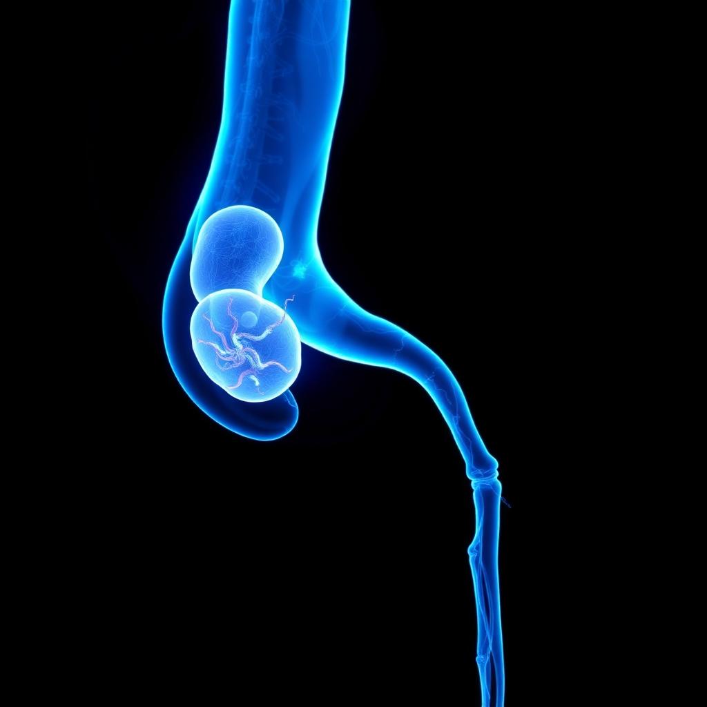
Plaque narrows the arteries of the leg until the muscle cannot get the oxygen it needs to move. It begins as pain after a set distance, and ends — if untreated — as pain at rest and tissue loss. The narrowing can be reopened from inside the artery.

Also known as
Peripheral arterial disease (PAD), also commonly described as poor blood circulation in the legs or blocked leg arteries, occurs when arteries supplying the legs become narrowed or blocked.
Poor blood circulation in the legs from peripheral artery disease is treated by angioplasty and stenting — the narrowed artery is opened from inside through a pinhole, restoring blood flow to the leg and foot.
How image-guided treatment helps
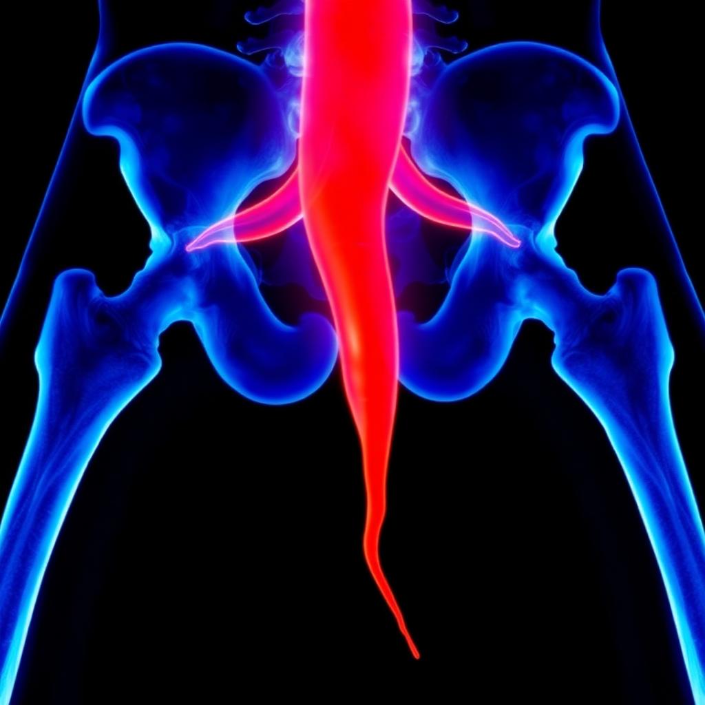
A wire is passed across the blockage under X-ray guidance, a balloon opens the artery, and a stent holds it open where needed. Inline flow to the foot is what allows rest pain to settle and wounds to heal.
Key facts
Cramping leg pain on walking, caused by narrowed leg arteries. Angioplasty and stenting reopen the artery through a puncture and restore walking distance.
- Common symptoms
- Cramping pain in the calf, thigh or buttock after walking a predictable distance · Pain that stops within minutes of standing still · Walking distance that shortens month by month
- Tests used
- Ankle-brachial index · Arterial Doppler ultrasound · Treadmill exercise test
- Treatment options
- Supervised exercise therapy · Smoking cessation · Statins and antiplatelets
- Recovery
- Procedure: Day 0: Angioplasty via puncture. Bed rest for a few hours, then walking. Most patients notice warmth returning. · Day 1: Discharged and walking. Puncture site checked and antiplatelet regimen started or adjusted.
02 · Symptoms
What patients notice.
- Cramping pain in the calf, thigh or buttock after walking a predictable distance
- Pain that stops within minutes of standing still
- Walking distance that shortens month by month
- Coldness or pallor of one foot
- Pain in the forefoot at night, eased by hanging the leg over the bed
- Loss of hair on the lower leg, shiny tight skin
- Slow-growing, thickened toenails
- Wounds on the foot that do not heal
- Erectile dysfunction, when the disease involves the aorto-iliac segment
03 · Causes
Why it happens.
- Atherosclerosis — cholesterol plaque building within the artery wall
- Thrombosis on an existing plaque, causing sudden worsening
- Embolism from the heart or a proximal aneurysm
- Arterial dissection
- Inflammatory arteritis, notably Buerger's disease in young smokers
- Radiation-induced arterial narrowing after previous treatment
04 · Risk factors
Who it affects.
- Smoking — the strongest modifiable risk factor for leg arteries
- Diabetes
- High blood pressure
- High cholesterol
- Age over 50
- Chronic kidney disease
- Known coronary or carotid artery disease
- Family history of vascular disease
05 · Warning signs
When to act immediately.
- Pain in the foot at rest or at night — the disease has progressed beyond exercise limitation
- Any non-healing wound or ulcer on the foot
- Blackening of a toe
- A suddenly cold, pale, painful and numb leg — an emergency requiring immediate assessment
- Walking distance falling rapidly over weeks rather than years
06 · Diagnosis
How it is confirmed.

- 01
History
Claudication distance, whether it is reproducible, rest pain, wounds, smoking history and cardiac symptoms.
- 02
Clinical examination
Palpation of femoral, popliteal, dorsalis pedis and posterior tibial pulses; skin temperature; capillary refill; auscultation for bruits.
- 03
Blood tests
Lipid profile, HbA1c, kidney function and haemoglobin — all shape both risk and the safety of contrast imaging.
- 04
Imaging
Ankle-brachial index and Doppler first; CT angiography to plan; catheter angiography when treatment is intended in the same sitting.
07 · Tests explained
Why each test is done.
Ankle-brachial index
Compares ankle to arm pressure. A simple number that confirms the diagnosis, grades severity and can be repeated to track progress.
Arterial Doppler ultrasound
Locates the level of narrowing and shows how the waveform changes across it, without radiation or contrast.
Treadmill exercise test
Used when resting pressures look normal but symptoms are typical. Pressures are measured after walking, when the deficit reveals itself.
CT angiography
A single scan mapping aorta to foot, showing calcification, length of blockage and run-off — the anatomy on which the treatment plan rests.
MR angiography
An alternative when kidney function or contrast allergy makes CT unsuitable.
Digital subtraction angiography
Performed in the catheter suite with the highest resolution for below-knee vessels, allowing diagnosis and treatment in one visit.
08 · Image-guided treatment options
Every route, stated plainly.
Supervised exercise therapy
Lifestyle
Structured walking to the point of pain, repeatedly, develops collateral flow. For mild claudication it rivals intervention.
Smoking cessation
Lifestyle
The single most effective measure. It changes both symptoms and the durability of any procedure performed.
Statins and antiplatelets
Medication
Reduce plaque progression and the risk of heart attack and stroke, which are the commonest causes of death in this disease.
Cilostazol
Medication
Improves walking distance in selected patients without heart failure.
Balloon angioplasty
Endovascular
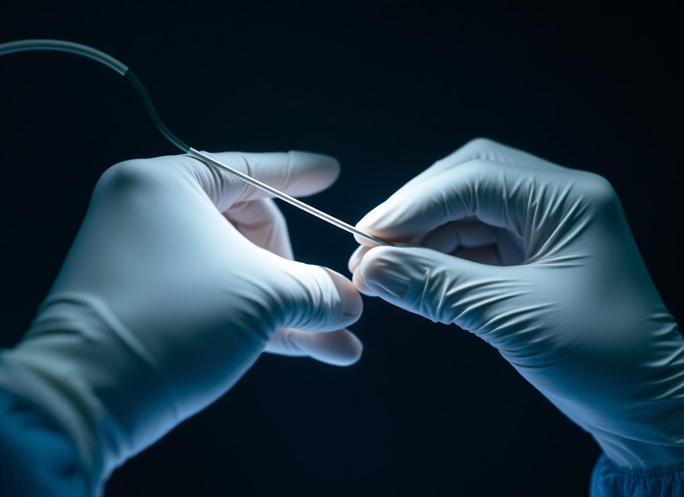
The narrowed segment is crossed with a wire and dilated with a balloon through a small puncture.
Stenting
Endovascular
A mesh scaffold holds the artery open where the vessel recoils or dissects after ballooning.
Drug-coated balloon
Endovascular
Delivers medication to the vessel wall to slow re-narrowing, particularly in the thigh artery.
Atherectomy
Endovascular
Removal or modification of heavily calcified plaque so the vessel can be dilated properly.
Bypass surgery
Surgery
Reserved for very long occlusions or after endovascular options have been exhausted.
09 · Evidence
What the imaging shows.
Narrowed segment
Flow restored
10 · The approach
How Dr. Mandeep treats it.
- 01
Claudication and limb-threatening ischemia are treated as different problems: the first is about quality of life, the second about saving a limb.
- 02
Every patient is assessed for heart and carotid disease at the same time — leg arteries rarely narrow in isolation.
- 03
Endovascular treatment is preferred first, preserving the vein for any future bypass.
- 04
Access is through a puncture at the groin, wrist or occasionally the foot artery, under local anaesthesia.
- 05
In long occlusions, both antegrade and retrograde approaches are used to cross the lesion.
- 06
Medical therapy is not optional background — statins, antiplatelets and smoking cessation are part of the procedure's success.
11 · Procedures
What is actually performed.
12 · Recovery
What the timeline looks like.

Procedure: Day 0
Angioplasty via puncture. Bed rest for a few hours, then walking. Most patients notice warmth returning.
Day 1
Discharged and walking. Puncture site checked and antiplatelet regimen started or adjusted.
1 Week
Return to light activity and routine lifestyle resume. Walking distance usually improves noticeably.
1 Month
Follow-up imaging and normal life. Ankle-brachial index repeated to document the gain.
This timeline is educational, not a personal recovery promise. Timing varies with the procedure, condition, other illnesses, and complications. Follow your treating team's discharge instructions.
13 · Prevention
What keeps it from returning.
- Stop smoking entirely
- Walk daily, pushing to near-maximal pain then resting and repeating
- Take statin and antiplatelet therapy without interruption
- Keep blood pressure and HbA1c at target
- Inspect the feet daily if you also have diabetes
- Report any fall in walking distance rather than adapting your life around it
- Maintain a healthy weight and a diet low in saturated fat
14 · Questions
22 questions patients ask.
What is peripheral arterial disease?+
Narrowing of the arteries supplying the limbs, usually by cholesterol plaque, reducing blood flow to the muscles and skin.
What is claudication?+
Reproducible cramping pain in the leg muscles brought on by walking a certain distance and relieved by rest. It is the classic symptom of PAD.
Is leg pain on walking always PAD?+
No. Spinal canal narrowing, arthritis and venous disease can mimic it. The pattern of relief and the ankle-brachial index usually separate them.
Will I lose my leg?+
Most people with claudication never do. The risk rises sharply once rest pain or tissue loss appears, which is why those symptoms need urgent assessment.
Can PAD be reversed?+
The plaque itself does not disappear, but flow can be restored by angioplasty and further progression slowed substantially by medication and stopping smoking.
Is walking safe when it causes pain?+
Yes, and it is therapeutic. Structured walking to the point of pain encourages collateral vessels to develop.
What is the ankle-brachial index?+
The ratio of ankle to arm blood pressure. Below 0.9 indicates arterial disease; below 0.4 indicates severe disease.
Why is my ABI normal if my arteries are calcified?+
In diabetes and kidney disease, calcified vessels resist compression and give falsely high readings. Toe pressures are used instead.
What does angioplasty involve?+
A needle puncture, a wire across the narrowing, and a balloon inflated to reopen the channel — sometimes followed by a stent. It is done awake, under local anaesthesia.
How long does a stent last?+
Many remain open for years. Durability depends on the vessel treated, lesion length, diabetes, kidney function and above all whether smoking stops.
What happens if the stent narrows again?+
Re-narrowing is usually treatable with a further balloon, often a drug-coated one. Surveillance is what allows it to be caught early.
Do I need surgery instead?+
Bypass is considered for very long blockages or when endovascular treatment fails. Most patients today are treated endovascularly first.
Why do I need a statin if my cholesterol is normal?+
Statins stabilise plaque and reduce heart attack and stroke risk in PAD independent of the starting cholesterol level.
Is PAD linked to heart disease?+
Strongly. The same process affects coronary and neck arteries, so cardiac and carotid assessment is part of good PAD care.
Can PAD cause erectile dysfunction?+
Yes, when the disease involves the aorto-iliac arteries. It can be the first symptom in some men.
What is critical limb ischemia?+
The advanced stage: pain at rest, ulceration or gangrene. It threatens the limb and requires prompt revascularisation.
How urgent is a suddenly cold, painful leg?+
An emergency. Acute limb ischemia has a window of a few hours before muscle and nerve damage becomes irreversible.
Does diabetes change the pattern of disease?+
Yes. Diabetic PAD tends to affect the smaller vessels below the knee, which requires different techniques and finer equipment.
Is contrast dye safe for my kidneys?+
Risk is managed with hydration, minimal contrast volume, and alternatives such as carbon dioxide angiography where kidney function is poor.
How often should I be followed up?+
Typically at one, three, six and twelve months after intervention, then yearly, with Doppler and pressure measurements each time.
What is the treatment for poor blood circulation in the legs?+
If leg arteries are narrowed or blocked, angioplasty with or without a stent reopens them through a pinhole puncture. Walking distance improves and non-healing wounds begin to close once blood flow returns.
Can leg artery blockage be treated without surgery?+
In most cases, yes. Angioplasty is performed through a small puncture in the groin or arm, usually with same-day or next-day discharge.
15 · Patient stories
Verified accounts only.
No testimonials are published here yet. Patient accounts will appear only once they are consented and verified — nothing on this page is written on a patient's behalf.
Patient stories →16 · Videos
See the procedure.
Animated films for this condition are being produced. In the meantime, the procedure pages above set out each step in sequence.