Condition
My wound isn't healing.
Diabetic foot
A wound on a diabetic foot is rarely only a wound. It is usually a circulation problem wearing the disguise of an infection. Restore the blood supply and tissue that was declared unsalvageable often heals.
Also known as
A diabetic foot wound may become difficult to heal when blood circulation to the foot is reduced — patients often search for a diabetic foot ulcer that will not heal.
A diabetic foot ulcer that will not heal often has reduced blood supply behind it. Restoring circulation by angioplasty gives the wound the blood flow it needs to close and can prevent amputation.
How image-guided treatment helps
Angiography maps the vessels below the knee and into the foot. Balloons open the diseased segments so that at least one straight line of flow reaches the wound.
Key facts
Non-healing foot ulcers in diabetes are usually a blood supply problem. Angioplasty below the knee restores flow and allows the wound to heal, avoiding amputation.
- Common symptoms
- A wound or ulcer that has not healed in weeks · Blackened skin at a toe tip or heel · Discharge, odour or surrounding redness
- Tests used
- Arterial Doppler ultrasound · Ankle-brachial and toe pressures · CT angiography
- Treatment options
- Below-knee angioplasty · Drug-coated balloon or stenting · Debridement
- Recovery
- Day 0: Angioplasty through a small puncture. The foot usually feels warmer within hours. Bed rest for a few hours, then walking. · Week 1: Debridement and dressing regimen begin against a now well-perfused wound bed. Antibiotics adjusted to culture.
02 · Symptoms
What patients notice.
- A wound or ulcer that has not healed in weeks
- Blackened skin at a toe tip or heel
- Discharge, odour or surrounding redness
- Numbness or a burning, tingling sensation in the feet
- Cold feet, or one foot colder than the other
- Pain in the foot at night, relieved by hanging the leg down
- Thin, shiny skin and loss of hair on the lower leg
- Slow-growing, thickened toenails
- A callus that has broken down at its centre
03 · Causes
Why it happens.
- Peripheral arterial disease — narrowed or blocked arteries below the knee reduce oxygen delivery to the tissue
- Diabetic neuropathy — loss of sensation, so injury goes unnoticed until it is deep
- Abnormal pressure loading from foot deformity, causing callus and then breakdown
- Infection entering through a small break in the skin
- Poor glycaemic control, which impairs both healing and immune response
- Ill-fitting footwear and walking barefoot
04 · Risk factors
Who it affects.
- Diabetes for more than ten years
- Poor blood sugar control
- Smoking
- Kidney disease or dialysis
- Previous ulcer or previous amputation
- Loss of protective sensation on testing
- Foot deformity — claw toes, Charcot foot, bunions
- Peripheral arterial disease
- Impaired vision, preventing daily self-examination
05 · Warning signs
When to act immediately.
- Any wound present for more than two weeks needs vascular assessment, not another dressing
- Blackening of a toe or heel — seek help the same day
- Fever, spreading redness or foul discharge suggests infection tracking deeper
- Sudden severe foot pain with pallor and coldness is an emergency
- An amputation recommended without a vascular study deserves a second opinion first
06 · Diagnosis
How it is confirmed.

- 01
History
How long the wound has been present, how it began, previous ulcers, smoking, dialysis, and whether there is rest pain at night.
- 02
Clinical examination
Pulses at the groin, knee, ankle and foot; wound depth probed to bone; sensation tested with a monofilament; the opposite foot always examined too.
- 03
Blood tests
HbA1c, inflammatory markers, kidney function and culture from the wound base rather than a surface swab.
- 04
Imaging
Doppler first, then CT or catheter angiography of the leg down to the pedal arch, because below-knee disease is where the answer usually lies.
07 · Tests explained
Why each test is done.
Arterial Doppler ultrasound
A painless first look at flow in the leg arteries. It shows where waveforms flatten out, pointing to the level of blockage.
Ankle-brachial and toe pressures
Numbers that predict healing. In diabetes ankle vessels are often calcified and falsely high, so toe pressure is the more honest measure.
CT angiography
Maps the whole arterial tree in one scan and plans the route a catheter will take before any puncture is made.
Digital subtraction angiography
The reference standard, done in the catheter suite. It shows the small vessels of the foot that other scans blur, and treatment can follow in the same sitting.
MRI of the foot
Answers whether infection has reached bone — osteomyelitis changes the treatment plan and the antibiotic duration.
Wound tissue culture
A deep tissue sample identifies the true organism. Surface swabs mostly grow contaminants and misdirect antibiotics.
08 · Image-guided treatment options
Every route, stated plainly.
Below-knee angioplasty
Endovascular
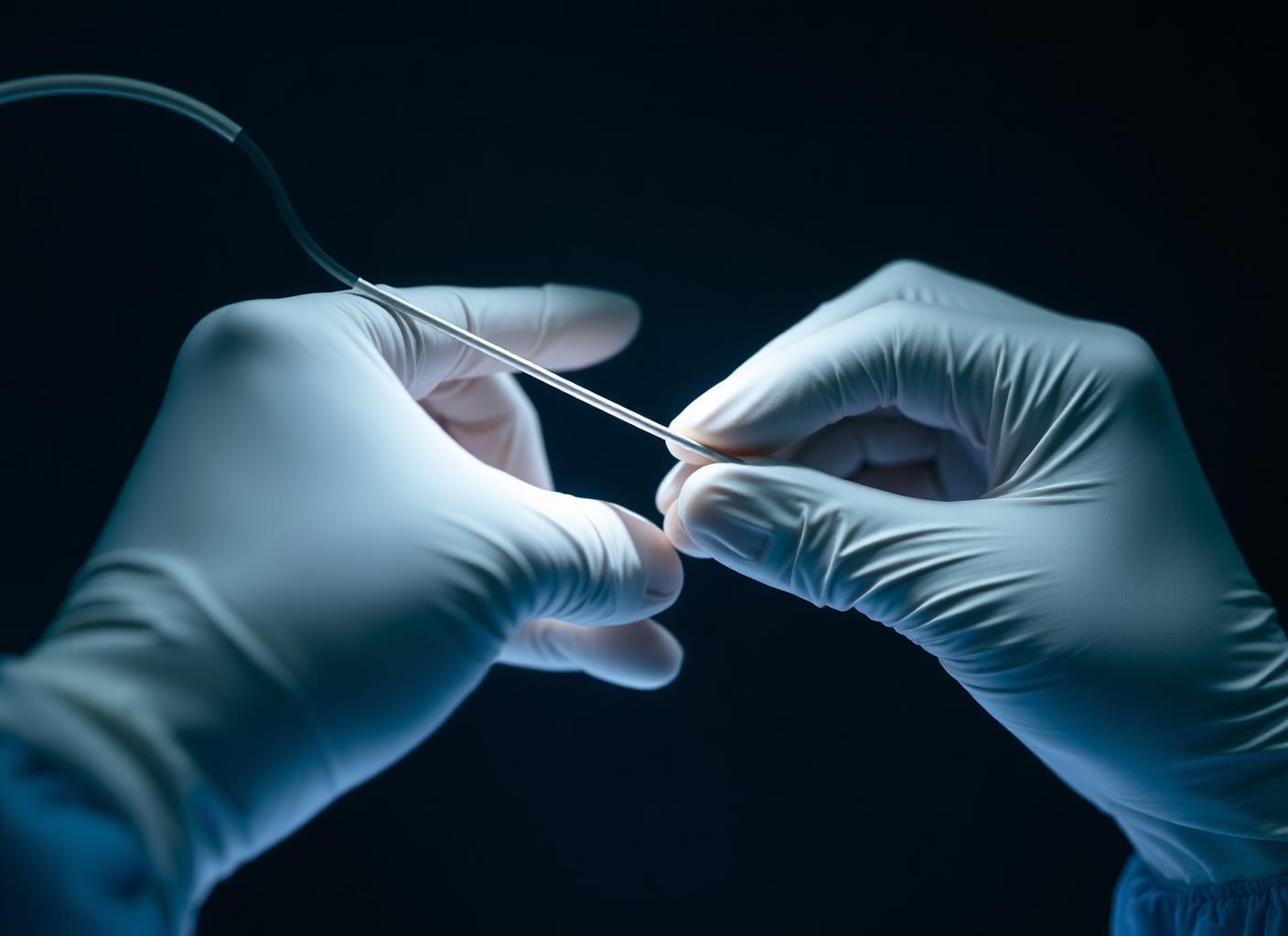
Balloon opening of the tibial and pedal arteries to bring direct flow to the wound bed. The central intervention in limb salvage.
Drug-coated balloon or stenting
Endovascular
Used above the knee where recoil or a long lesion makes plain ballooning insufficient.
Debridement
Surgery
Removal of dead tissue so the wound has a living edge to heal from. Repeated as needed, timed after flow is restored.
Targeted antibiotics
Medication
Guided by deep culture. Antibiotics cannot reach tissue that has no blood supply, which is why circulation is addressed first.
Offloading
Lifestyle
Total contact casting or offloading footwear removes pressure from the ulcer. Healing frequently fails for want of this alone.
Glycaemic and nutritional control
Lifestyle
Sugar control, protein intake and smoking cessation change healing rates measurably.
Minor amputation
Surgery
Removal of a single non-viable toe or ray, performed after revascularisation so the wound can close — a limb-preserving operation, not a failure.
09 · Evidence
What the imaging shows.
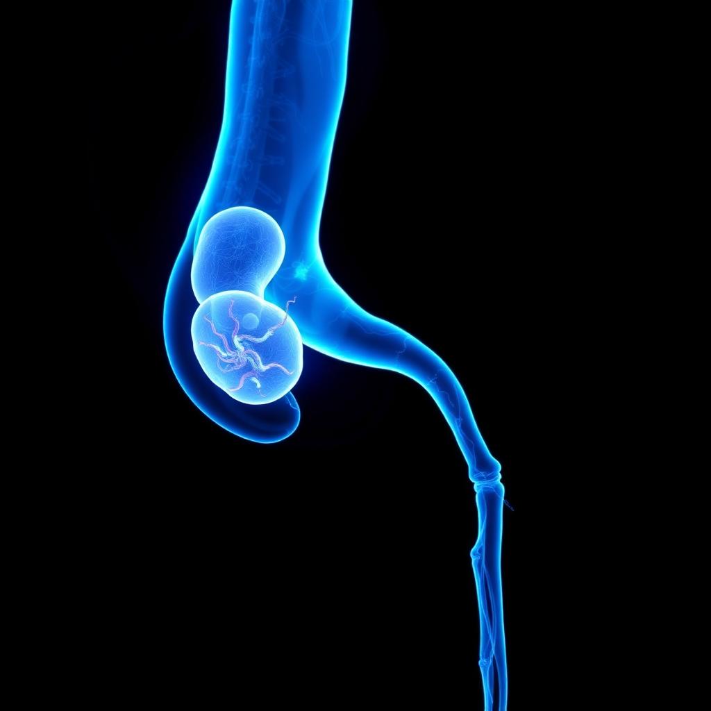
Below-knee vessel blocked
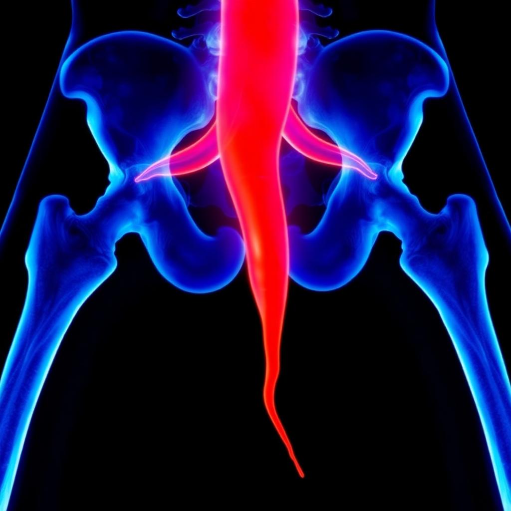
Below-knee flow re-established
10 · The approach
How Dr. Mandeep treats it.
- 01
The first question is always circulation. No wound care plan is agreed before the arteries below the knee have been imaged.
- 02
The target is direct flow to the angiosome — the specific territory of the foot in which the wound sits — not merely an improved pulse.
- 03
Below-knee vessels are treated to the pedal arch where possible, using long balloons and low-profile wires.
- 04
Procedures are done through a puncture, usually under local anaesthesia, and most patients go home the same or next day.
- 05
Wound care, offloading, podiatry and diabetes control run in parallel; revascularisation alone does not close a wound.
- 06
Amputation is considered only after the arterial tree has been assessed and treated.
11 · Procedures
What is actually performed.
12 · Recovery
What the timeline looks like.

Day 0
Angioplasty through a small puncture. The foot usually feels warmer within hours. Bed rest for a few hours, then walking.
Week 1
Debridement and dressing regimen begin against a now well-perfused wound bed. Antibiotics adjusted to culture.
Weeks 2 to 6
Granulation tissue appears and the wound edge contracts. Offloading maintained without exception.
Months 2 to 4
Most ulcers close in this window when flow has been restored and pressure removed.
Month 6
Doppler review to confirm the treated segment remains open, and footwear reassessed.
Long term
Daily foot inspection, annual vascular review, and immediate reporting of any new break in the skin.
This timeline is educational, not a personal recovery promise. Timing varies with the procedure, condition, other illnesses, and complications. Follow your treating team's discharge instructions.
13 · Prevention
What keeps it from returning.
- Inspect both feet daily, including between the toes and the soles, using a mirror if needed
- Never walk barefoot, indoors or outdoors
- Have calluses trimmed by a podiatrist, not at home
- Wear properly fitted footwear and check inside shoes before wearing
- Keep HbA1c to the target agreed with your physician
- Stop smoking
- Have pulses and sensation checked at least once a year
- Report any new wound within days, not weeks
14 · Questions
22 questions patients ask.
Why won't my diabetic foot wound heal?+
Most often because the arteries supplying the foot are narrowed and the tissue is not receiving enough oxygen. Infection and pressure are common additional factors.
Can my foot be saved?+
In a large proportion of cases yes, if the blood supply can be restored before tissue loss becomes extensive. That question should be answered with an angiogram, not an opinion.
Is amputation the only option?+
It should be the last option. Below-knee angioplasty has changed what is salvageable, and a full vascular assessment should precede any amputation decision.
What is angioplasty in diabetic foot?+
A thin balloon is passed through a needle puncture into the leg arteries and inflated to reopen the blocked segments, restoring direct flow to the wound.
Is angioplasty painful?+
It is performed under local anaesthesia. Patients feel pressure rather than pain, and often report warmth in the foot during the case.
How long does the procedure take?+
Typically one to two hours, depending on how many vessels are blocked and how long the blockages are.
Will I be admitted?+
Usually one night, sometimes as a day case. Wound and infection status often decides this rather than the procedure itself.
What is the difference between poor circulation and infection?+
Infection produces redness, warmth, discharge and fever. Poor circulation produces coldness, pain at rest, blackening and a wound that simply does not progress. They frequently coexist.
Why is a Doppler done first?+
It is quick, painless and identifies whether there is arterial disease at all, and roughly at what level, before contrast studies are considered.
Why do I need CT angiography as well?+
Doppler shows there is a problem; CT angiography shows the exact anatomy needed to plan a route for the catheter.
Is contrast safe if my kidneys are weak?+
Contrast is used cautiously with hydration, minimal volume and sometimes carbon dioxide angiography instead. Kidney function is always checked first.
Can antibiotics alone heal the ulcer?+
Not if blood supply is inadequate — antibiotics travel in blood and cannot reach tissue that has none. Circulation must be addressed for antibiotics to work.
How long will the wound take to heal after angioplasty?+
Commonly six to sixteen weeks, depending on wound size, depth, infection and how consistently pressure is kept off it.
Will the artery block again?+
It can, particularly in diabetes and kidney disease. Follow-up Doppler, medication and risk factor control reduce the chance, and a repeat procedure is possible if needed.
Does smoking really matter at this stage?+
Enormously. Continued smoking is one of the strongest predictors of failed healing and repeat blockage.
What is offloading and why does it matter?+
It means removing weight from the ulcer with a cast or special footwear. A wound that is walked on each day cannot close, however good the circulation.
What is Charcot foot?+
Progressive collapse of the bones of the foot due to neuropathy, changing its shape and creating new pressure points. It requires immobilisation and specialist care.
Is a black toe always lost?+
Not always. If flow is restored early, the demarcation can stay limited and only a small portion may need removal.
How often should I have my feet checked?+
At least annually if you have no problems, and far more often if you have neuropathy, arterial disease or a previous ulcer.
Can this happen to my other foot?+
Yes. The disease process is systemic, so the opposite foot is examined and monitored as a matter of routine.
Why is my diabetic foot wound not healing?+
A wound cannot heal without blood supply. In diabetes, the small arteries below the knee are often narrowed, so the wound is starved even when dressings and antibiotics are correct.
Can amputation be avoided in diabetic foot?+
Often, yes — if blood flow is restored early and infection is controlled. The decision depends on how much tissue is still viable.
15 · Patient stories
Verified accounts only.
No testimonials are published here yet. Patient accounts will appear only once they are consented and verified — nothing on this page is written on a patient's behalf.
Patient stories →16 · Videos
See the procedure.
Animated films for this condition are being produced. In the meantime, the procedure pages above set out each step in sequence.