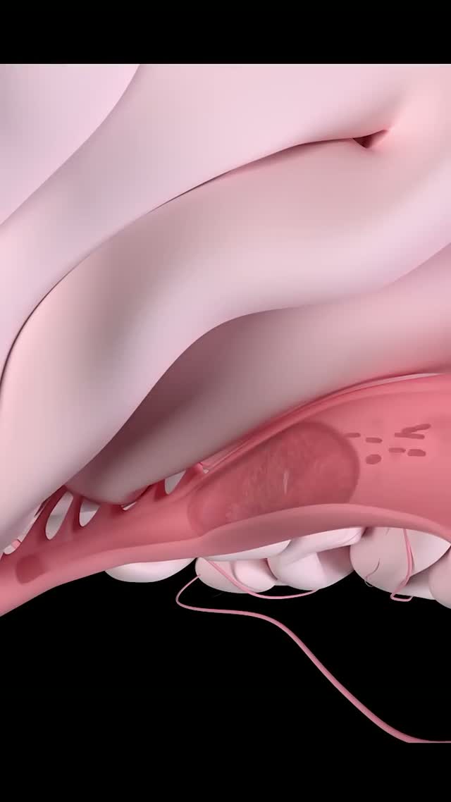
Condition
One leg swelled up overnight.
Deep vein thrombosis
A clot in the deep veins of the leg blocks the route blood takes back to the heart. The immediate danger is that part of it travels to the lungs. The long-term danger is a leg left permanently swollen and heavy — which early clot removal can prevent.
Also known as
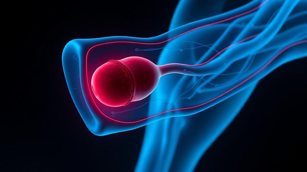
Deep vein thrombosis (DVT) is a blood clot forming in a deep vein, usually of the leg, causing swelling and pain.
Deep vein thrombosis is treated with blood thinners, and in selected patients with extensive clot, by catheter-directed thrombolysis or clot removal to protect the vein valves.
How image-guided treatment helps
A catheter is placed into the clotted vein under ultrasound and X-ray guidance so that clot-dissolving medicine acts directly where the clot is, or the clot is aspirated out.
Key facts
Sudden swelling and pain in one leg from a deep vein clot. Anticoagulation prevents extension; catheter-directed clot removal protects the valves in selected patients.
- Common symptoms
- Swelling in one leg or arm · Pain or tenderness when standing or walking · Warmth in the swollen or painful area
- Tests used
- Compression duplex ultrasound · D-dimer · CT venography
- Treatment options
- Anticoagulation · Catheter-directed thrombolysis · Pharmacomechanical thrombectomy
- Recovery
- Day 0: Anticoagulation begins immediately once diagnosis is confirmed. Walking is encouraged, not restricted. · Days 1 to 3: Where intervention is indicated, catheter-directed lysis is performed and the vein reassessed daily.
02 · Symptoms
What patients notice.
- Swelling in one leg or arm
- Pain or tenderness when standing or walking
- Warmth in the swollen or painful area
- Red or discolored skin
- Sudden breathlessness or chest pain
03 · Causes
Why it happens.
- Sitting still for a long time (like on a long flight or car ride)
- Long periods of bed rest or inactivity after surgery or illness
- Injury or damage to a deep vein
- Medical conditions like cancer or inherited clotting disorders
- Pregnancy or taking birth control pills / hormone therapy
04 · Risk factors
Who it affects.
- Previous DVT or pulmonary embolism
- Family history of clotting
- Age above 60
- Obesity
- Smoking
- Active malignancy
- Dehydration
05 · Warning signs
When to act immediately.
- Pulmonary embolism: Sudden breathlessness, chest pain on breathing, coughing blood or fainting — this is a life-threatening emergency.
- Venous thromboembolism (VTE): The collective term for DVT and PE.
- A piece of the blood clot can break off, travel through the bloodstream, and block a blood vessel in the lungs.
06 · Diagnosis
How it is confirmed.

- 01
History
Speed of onset, travel, surgery, immobility, hormone use, cancer, previous clots and family history.
- 02
Clinical examination
Circumference measured and compared with the other leg, tenderness along the deep veins, warmth, and assessment for breathlessness.
- 03
Blood tests
D-dimer to exclude clot in low-risk patients, plus full blood count, kidney function and clotting profile before anticoagulation.
- 04
Imaging
Compression duplex ultrasound confirms the clot and its extent; CT venography assesses the pelvic veins and CT pulmonary angiography the lungs.
07 · Tests explained
Why each test is done.
Compression duplex ultrasound
The main test. A vein that does not compress under the probe contains clot. It also shows how far up the clot extends, which changes management.
D-dimer
Highly sensitive but not specific. A normal result in a low-risk patient safely excludes DVT; a raised one only means imaging is needed.
CT venography
Looks above the groin, where ultrasound is unreliable, and identifies iliac vein compression as an underlying cause.
CT pulmonary angiography
Performed when there is breathlessness or chest pain, to confirm or exclude pulmonary embolism.
Thrombophilia screen
Considered in young patients, recurrent clots or a strong family history — the result may determine how long anticoagulation continues.
Cancer screening
An unprovoked clot in an older patient warrants age-appropriate cancer assessment, as it can be the first sign.
08 · Image-guided treatment options
Every route, stated plainly.

Anticoagulation
Medication
The foundation of treatment. It prevents the clot extending and embolising while the body dissolves it, but it does not itself remove clot.
Catheter-directed thrombolysis
Endovascular
Clot-dissolving drug delivered directly into the clot through a catheter over hours, used in extensive iliofemoral clot to preserve valves.
Pharmacomechanical thrombectomy
Endovascular
Combined drug and mechanical clot removal in a single session, reducing drug dose and treatment time.
Venous stenting
Endovascular
Used when an underlying iliac vein compression is found, to keep the vein open and prevent recurrence.
IVC filter
Endovascular
A temporary filter in the vena cava for patients who cannot receive anticoagulation, retrieved as soon as it is safe.
Graduated compression
Lifestyle
Reduces swelling and symptoms during recovery and helps limit post-thrombotic change.
Early mobilisation
Lifestyle
Walking is encouraged once anticoagulation is started — bed rest does not reduce embolism risk.
09 · Evidence
What the imaging shows.
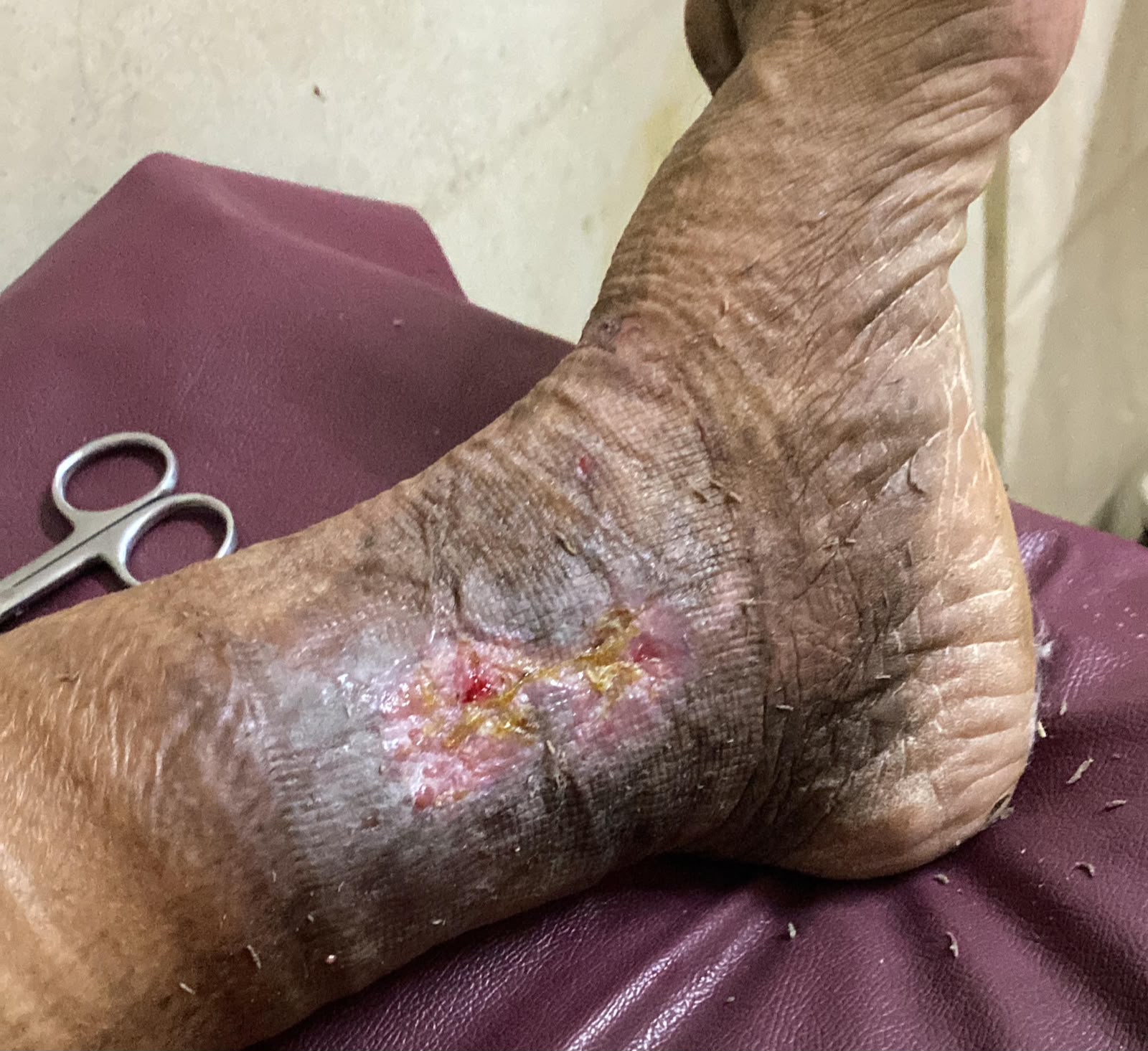
Thrombosed vein
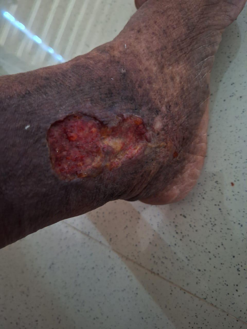
Vein recanalised
10 · The approach
How Dr. Mandeep treats it.
- 01
The first decision is anatomical: clot confined below the knee behaves very differently from clot filling the iliac vein.
- 02
Extensive iliofemoral clot in a younger, active patient is considered for catheter-directed removal, because anticoagulation alone often leaves a damaged, swollen leg.
- 03
Any patient with left-sided iliofemoral DVT is assessed for iliac vein compression, which is otherwise missed and causes recurrence.
- 04
Filters are used sparingly, always with a retrieval plan and date.
- 05
The cause is pursued in every unprovoked case — duration of anticoagulation depends on it.
- 06
Follow-up includes surveillance for post-thrombotic syndrome, which is preventable and often ignored.
11 · Procedures
What is actually performed.
12 · Recovery
What the timeline looks like.

Day 0
Anticoagulation begins immediately once diagnosis is confirmed. Walking is encouraged, not restricted.
Days 1 to 3
Where intervention is indicated, catheter-directed lysis is performed and the vein reassessed daily.
Week 1 to 2
Swelling begins to settle. Compression stocking worn during the day.
Month 1
Review of symptoms, bleeding risk and adherence. Duplex to document the residual clot burden.
Months 3 to 6
Decision on whether anticoagulation continues, based on whether the clot was provoked and on test results.
Year 1 and beyond
Monitoring for post-thrombotic syndrome — persistent swelling, heaviness or skin change — which is treatable if identified.
This timeline is educational, not a personal recovery promise. Timing varies with the procedure, condition, other illnesses, and complications. Follow your treating team's discharge instructions.
13 · Prevention
What keeps it from returning.
- Walk and flex the ankles hourly during long flights or drives
- Stay well hydrated when travelling
- Use prescribed prophylaxis after surgery or during hospital admission
- Wear compression stockings if you have had a previous clot
- Discuss clot risk before starting oestrogen-containing contraception
- Maintain a healthy weight and stop smoking
- Report one-sided leg swelling promptly rather than waiting to see if it settles
14 · Questions
23 questions patients ask.
What is deep vein thrombosis?+
A blood clot forming in the deep veins, usually of the leg, obstructing the return of blood to the heart.
How do I know if leg swelling is a clot?+
Clot typically affects one leg, develops over hours to days, and is accompanied by calf tightness or pain. Only an ultrasound can confirm it.
Is DVT dangerous?+
Yes. Part of the clot can travel to the lungs and cause a pulmonary embolism, which can be fatal. It is treatable when caught early.
What is a pulmonary embolism?+
A clot that has travelled to the lung arteries, causing sudden breathlessness, chest pain on breathing or collapse. It requires emergency care.
How is DVT diagnosed?+
Compression duplex ultrasound is the standard test. D-dimer helps exclude it in low-risk patients.
Can I walk with a DVT?+
Yes, once anticoagulation has started. Walking helps and does not increase the risk of embolism.
Should I massage the leg?+
No. Avoid massage over an acute clot.
How long will I be on blood thinners?+
At least three months. Longer or indefinitely if the clot was unprovoked, recurrent, or associated with cancer or a clotting disorder.
Do blood thinners dissolve the clot?+
They prevent it growing and travelling while the body's own system breaks it down. They do not directly dissolve it.
What is catheter-directed thrombolysis?+
A catheter is placed into the clot and a clot-dissolving drug infused directly, removing clot far faster and helping preserve the vein valves.
Who benefits from clot removal rather than tablets alone?+
Typically younger, active patients with extensive clot in the thigh and pelvic veins, treated within about two weeks of onset.
What is post-thrombotic syndrome?+
Chronic swelling, heaviness, discoloration and sometimes ulceration after a DVT, caused by damaged valves and residual obstruction.
Can DVT come back?+
Yes, particularly after unprovoked clots. Recurrence risk guides how long anticoagulation continues.
What is venous thromboembolism (VTE)?+
VTE is the collective term for DVT and pulmonary embolism (PE).
Can a clot travel to the lungs?+
Yes. This is called a pulmonary embolism and is the primary danger of DVT.
What is May-Thurner syndrome?+
Compression of the left iliac vein by the overlying right iliac artery, predisposing to left leg DVT. It is treated by stenting the vein.
Is flying safe after a DVT?+
Usually, once established on anticoagulation, with compression stockings, hydration and regular movement. Check with your physician first.
Does DVT happen in the arm?+
Yes, most often around central lines and pacemaker leads, or in athletes with thoracic outlet compression.
Should my family be tested?+
Testing is considered when there is a strong family history or clots at a young age. It is not routine for everyone.
Can I take contraceptive pills after a DVT?+
Oestrogen-containing pills are generally avoided afterwards. Alternative methods are discussed with your gynaecologist.
What is an IVC filter?+
A small device placed in the main abdominal vein to catch clots, used when blood thinners cannot be given. It should be removed once it is no longer needed.
Will my leg return to normal size?+
Most legs improve substantially over weeks to months. Compression and, in selected cases, early clot removal improve the final result.
What are the signs of a blood clot in the leg?+
Sudden swelling of one leg, calf pain or tightness, warmth and redness. Breathlessness or chest pain alongside these is an emergency.
15 · Patient stories
Verified accounts only.
No testimonials are published here yet. Patient accounts will appear only once they are consented and verified — nothing on this page is written on a patient's behalf.
Patient stories →16 · Videos