Condition
The worst headache of my life.
Brain aneurysm
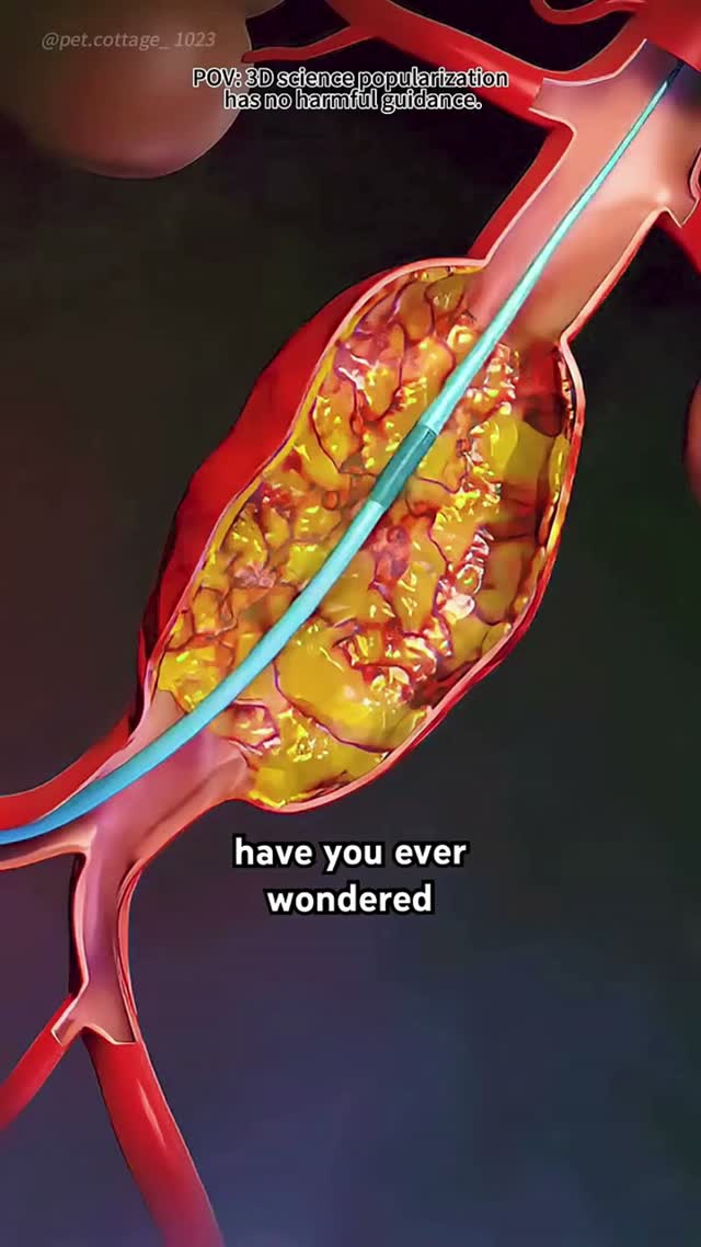
An aneurysm is a weak spot on a brain artery that balloons outward. Most are silent. The ones that tear announce themselves with a headache unlike any other. Both can be treated from inside the vessel, filling the sac with fine coils until blood no longer enters it.

Also known as
A brain (cerebral) aneurysm is a weak, ballooned area in the wall of a blood vessel supplying the brain.
A brain aneurysm can often be treated from inside the blood vessel by endovascular coiling or flow diversion, sealing the aneurysm without opening the skull.
How image-guided treatment helps
A micro-catheter is navigated into the aneurysm sac and soft platinum coils are packed inside it, or a flow-diverting stent is placed across its neck. Blood stops entering the weak pouch and it clots off safely.
Key facts
A ballooned weak point on a brain artery. Endovascular coiling or flow diversion seals it from inside the vessel, without opening the skull.
- Common symptoms
- Sudden, explosive headache reaching maximum intensity within seconds — the hallmark of rupture · Neck stiffness and intolerance of light · Vomiting with the headache
- Tests used
- Non-contrast CT head · CT angiography · Lumbar puncture
- Treatment options
- Endovascular coiling · Balloon or stent-assisted coiling · Flow diversion
- Recovery
- Day 0: After coiling, care continues in intensive care for ruptured cases, with blood pressure control and monitoring of conscious level. · Days 1 to 3: Unruptured patients are usually discharged within a day or two. Ruptured patients remain monitored for vasospasm.
02 · Symptoms
What patients notice.
- Sudden, explosive headache reaching maximum intensity within seconds — the hallmark of rupture
- Neck stiffness and intolerance of light
- Vomiting with the headache
- Brief loss of consciousness or collapse
- Double vision or a drooping eyelid, from pressure on the nerve to the eye
- A dilated pupil on one side
- Seizure
- Most unruptured aneurysms cause no symptoms at all and are found incidentally
03 · Causes
Why it happens.
- Weakness in the arterial wall at branch points, where flow stress is highest
- Long-standing high blood pressure
- Smoking, which both weakens the wall and increases rupture risk
- Inherited connective tissue disorders such as Ehlers-Danlos or Marfan syndrome
- Autosomal dominant polycystic kidney disease
- Arterial dissection or trauma
- Infection of the vessel wall, producing a mycotic aneurysm
04 · Risk factors
Who it affects.
- Smoking
- High blood pressure
- Family history of aneurysm or subarachnoid haemorrhage
- Polycystic kidney disease
- Female sex, particularly after menopause
- Age above 40
- Heavy alcohol use
- Cocaine or stimulant use
05 · Warning signs
When to act immediately.
- A sudden severe headache unlike any previous headache — call emergency services immediately
- Headache with neck stiffness, vomiting or light intolerance
- A new drooping eyelid or double vision, which can precede rupture
- A brief severe headache days earlier — a sentinel bleed that must not be dismissed
- Any collapse or seizure with headache
06 · Diagnosis
How it is confirmed.

- 01
History
The speed of headache onset is the critical detail. Thunderclap onset means subarachnoid haemorrhage until proven otherwise.
- 02
Clinical examination
Conscious level, pupil size and reaction, eye movements, neck stiffness and focal neurological deficit are documented and graded.
- 03
Blood tests
Full blood count, clotting, electrolytes and kidney function, both for baseline and to prepare for contrast and intervention.
- 04
Imaging
Urgent CT of the head, then CT angiography. Lumbar puncture if CT is negative but suspicion remains, and catheter angiography for definitive anatomy.
07 · Tests explained
Why each test is done.
Non-contrast CT head
Detects blood in the subarachnoid space with very high sensitivity in the first hours. It is the immediate test in any thunderclap headache.
CT angiography
Locates the aneurysm, measures its neck and dome, and shows the surrounding branches — the information that decides coiling versus clipping.
Lumbar puncture
Performed when CT is negative but the history is convincing. Blood breakdown pigment in the fluid confirms a bleed that CT has missed.
Digital subtraction angiography
The reference standard, with 3D rotational imaging. It shows small aneurysms other studies miss and defines the working projection for treatment.
MR angiography
Used for screening and for follow-up surveillance without radiation or iodinated contrast.
Transcranial Doppler
Monitors for vasospasm in the days after a bleed, when arteries can narrow and cause a delayed stroke.
08 · Image-guided treatment options
Every route, stated plainly.

Endovascular coiling
Endovascular
Fine platinum coils are packed into the aneurysm sac through a microcatheter until blood no longer enters it and the sac clots off.
Balloon or stent-assisted coiling
Endovascular
Used for wide-necked aneurysms, where a balloon or stent holds the coils inside the sac and protects the parent artery.
Flow diversion
Endovascular
A dense mesh stent placed across the neck redirects flow past the aneurysm, which thromboses and shrinks over months. Used for large or complex aneurysms.
Surgical clipping
Surgery
A titanium clip placed across the neck through a craniotomy. Preferred for some anatomy, particularly wide-necked middle cerebral aneurysms.
Blood pressure control
Medication
Fundamental both to prevent rupture and to manage the acute phase after a bleed.
Nimodipine
Medication
Given after subarachnoid haemorrhage to reduce the risk of delayed ischemic injury from vasospasm.
Surveillance
Observation
Small, low-risk unruptured aneurysms may be monitored with interval imaging rather than treated.
09 · Evidence
What the imaging shows.
Imaging figures for this condition are being prepared. Case imaging is published only once it is anonymised and verified.
10 · The approach
How Dr. Mandeep treats it.
- 01
A ruptured aneurysm is secured as early as possible — the highest risk of a second bleed is within the first days.
- 02
Anatomy decides technique. Neck width, dome-to-neck ratio, branch involvement and vessel tortuosity are assessed on 3D angiography before a device is chosen.
- 03
Access is through the wrist or groin. The skull is not opened, and there is no brain retraction.
- 04
Wide-necked and complex aneurysms are treated with balloon assistance, stent assistance or flow diversion rather than forced into simple coiling.
- 05
Unruptured aneurysms are discussed honestly: size, location, growth and patient age are weighed against the risk of treatment, and observation is a legitimate outcome of that discussion.
- 06
Follow-up imaging is scheduled from the outset, because coil compaction and recurrence must be detected early.
11 · Procedures
What is actually performed.
- →
Cerebral aneurysm coiling
Packing an aneurysm until blood no longer enters it.
Embolization
Performed through a small puncture under image guidance — discussed in detail at consultation.
12 · Recovery
What the timeline looks like.

Day 0
After coiling, care continues in intensive care for ruptured cases, with blood pressure control and monitoring of conscious level.
Days 1 to 3
Unruptured patients are usually discharged within a day or two. Ruptured patients remain monitored for vasospasm.
Days 4 to 14
The vasospasm window after a bleed, monitored with Doppler and treated promptly if it develops.
Weeks 2 to 6
Gradual return to activity for unruptured cases. After a bleed, fatigue and headache commonly persist for weeks.
Month 3 to 6
First follow-up angiography or MR angiography to confirm the aneurysm remains occluded.
Year 1 and beyond
Ongoing surveillance imaging at intervals, with lifelong blood pressure control and no smoking.
This timeline is educational, not a personal recovery promise. Timing varies with the procedure, condition, other illnesses, and complications. Follow your treating team's discharge instructions.
13 · Prevention
What keeps it from returning.
- Stop smoking completely — the single strongest modifiable factor
- Keep blood pressure consistently controlled
- Avoid stimulant drugs
- Limit alcohol
- Consider screening if two or more first-degree relatives have had an aneurysm
- Screening is advised in polycystic kidney disease
- Attend scheduled surveillance scans for known untreated aneurysms
14 · Questions
22 questions patients ask.
What is a brain aneurysm?+
A weakened, ballooned area on the wall of a brain artery. It may remain stable for life or, less commonly, tear and bleed.
Will my aneurysm burst?+
Most do not. Risk depends on size, location, shape, growth on serial imaging, smoking, blood pressure and family history.
What does a rupture feel like?+
A sudden, explosive headache reaching maximum severity within seconds, often with vomiting, neck stiffness and light intolerance.
What is coiling?+
Soft platinum coils are delivered through a microcatheter into the aneurysm until it is packed and blood can no longer enter. The skull is not opened.
Coiling or clipping — which is better?+
Neither is universally better. Coiling avoids open surgery and generally has a faster recovery; clipping suits certain anatomy. The decision is made on the angiogram.
What is a flow diverter?+
A fine-mesh stent placed in the parent artery across the aneurysm neck. It redirects flow so the aneurysm clots and shrinks over months.
How long does coiling take?+
Typically one to three hours depending on the complexity of the anatomy.
Is coiling done under general anaesthesia?+
Yes, usually, so the patient is completely still during precise work inside small vessels.
What are the risks?+
Rupture during the procedure, clot formation causing stroke, coil migration and access site complications. These are weighed against the risk of leaving the aneurysm untreated.
Will I need lifelong follow-up?+
Yes, with interval imaging. Coils can compact and aneurysms occasionally recur, and new aneurysms can form.
Can an aneurysm come back after coiling?+
A minority recur, particularly larger and wide-necked ones. That is why surveillance imaging is scheduled.
Do I need blood thinners after treatment?+
After stent-assisted coiling or flow diversion, dual antiplatelet therapy is required for a period. Simple coiling usually does not need it.
Should my family be screened?+
Screening is generally offered when two or more first-degree relatives have had an aneurysm, or with polycystic kidney disease.
Can I exercise with an untreated aneurysm?+
Moderate exercise is generally acceptable; heavy straining and extreme exertion are usually discouraged. Advice is individual.
Does stress cause rupture?+
Rupture often occurs during ordinary activity. Sustained high blood pressure matters far more than momentary stress.
What is a sentinel headache?+
A brief severe headache days or weeks before a major bleed, caused by a small leak. It is an urgent warning and frequently missed.
What is vasospasm?+
Narrowing of brain arteries several days after a bleed, which can cause a delayed stroke. It is monitored for and treated actively.
Can I fly after coiling?+
Usually yes, once recovered and cleared at follow-up. Timing depends on whether there was a bleed.
Will an MRI be safe with coils in place?+
Modern coils and stents are MRI-compatible at standard field strengths. Always inform the radiographer.
Can small aneurysms just be watched?+
Yes. Many small, stable aneurysms in low-risk locations are monitored with imaging rather than treated.
Can a brain aneurysm be treated without open surgery?+
Yes. Most aneurysms suitable for treatment can be coiled or flow-diverted from inside the blood vessel through a pinhole in the wrist or groin artery.
What are the warning signs of a brain aneurysm?+
Many are silent. A sudden, severe headache unlike any before, neck stiffness, double vision or a drooping eyelid needs urgent imaging.
15 · Patient stories
Verified accounts only.
No testimonials are published here yet. Patient accounts will appear only once they are consented and verified — nothing on this page is written on a patient's behalf.
Patient stories →16 · Videos